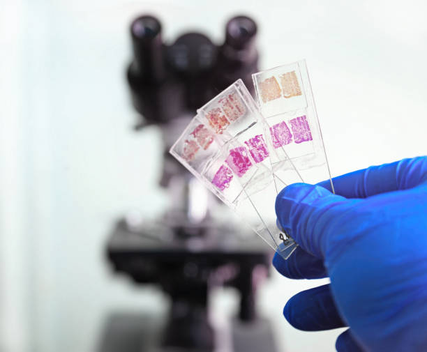
Glass slides in the laboratory. Hand in blue glove holding glass organ samples. Histological examination. The microscope in the background blurred. Pathologist at work.
Browse 3,000+ biopsy slide stock photos and images available, or start a new search to explore more stock photos and images.

Glass slides in the laboratory. Hand in blue glove holding glass organ samples. Histological examination. The microscope in the background blurred. Pathologist at work.
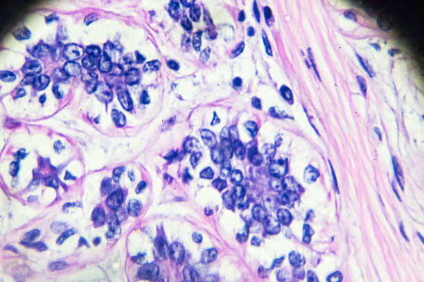
Breast Cancer under light microscopy zoom in different areas
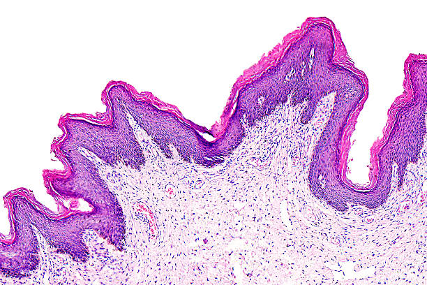
Skin papilloma of a human, highly detailed segment of panorama. Photomicrograph as seen under the microscope, 10x zoom.

Close view of Histopathology slides stained with hematoxylin and eosin or HE stain, ready for microscopic examination with Laboratory background, Histology.
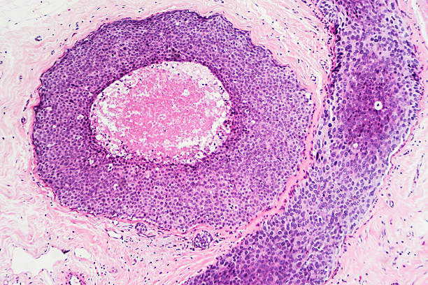
Breast cancer - ductal carcinoma in situ (DCIS): Tumor cells are confined to the mammary ducts. No invasion is seen (photographed and uploaded by US board certified surgical pathologist).
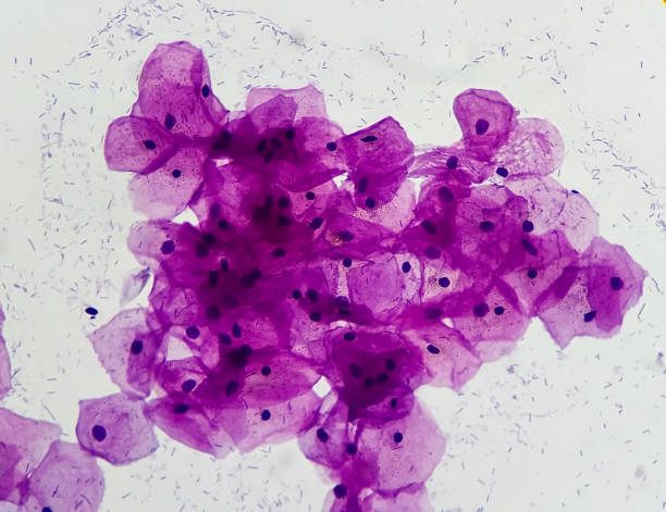
Pap's smear Microscopic 100x show human cervical epithelial cells . To diagnosis woman cervix cancer cells at medical laboratory.
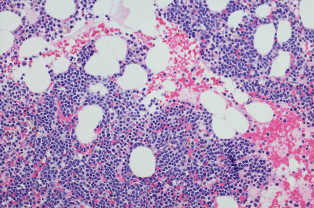
Micrograph of myeloma neoplasm bone marrow biopsy. Hematoxylin and eosin staining (H&E)

Female Scientist researcher using microscope in laboratory. Medical healthcare technology and pharmaceutical research and development concept.

Laboratory assistant works with microscope at the modern laboratory

Slide glass with histological sections for microscopy

Archival Biopsy Test Kit 3D Isometric Set. Laboratory Equipment for Medical Analysis or Scientific Oncology Translational Research. Labelled Specimen Slides Tumor Block Liquid Biposy Box

Immunohistochemistry staining is used in histopathology to do research or diagnosis.

Blurred background for histopathology-related presentation. Stained histological tissue samples on glass, fixed tissue, automatic pipette and other work-related tools. Toned image, text space.
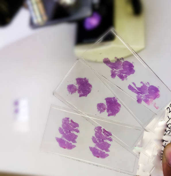
Close view of Histopathology slides stained with hematoxylin and eosin or HE stain, ready for microscopic examination with Laboratory background, Histology.
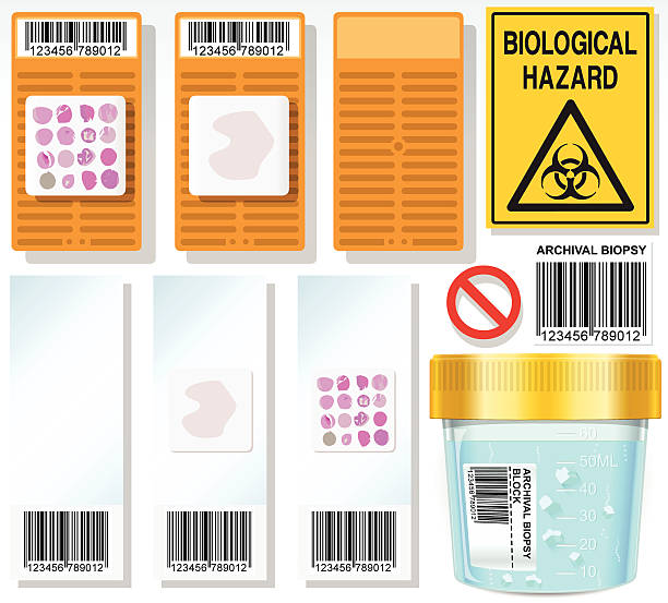
Detailed illustration of a Archival Biopsy Complete Set This illustration is saved in EPS10 with color space in RGB.
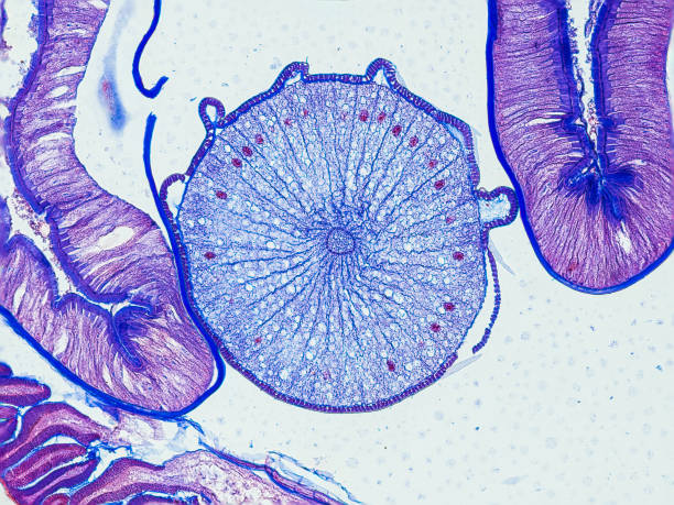
ascaris megalocephala cross section under the microscope showing an ovary - optical microscope x200 magnification
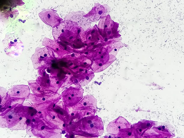
Squamous epithelial cells. Micrograph close up image at medical laboratory.

Blurred background for histopathology-related presentation. Stained histological tissue samples on glass, fixed tissue, automatic pipette and other work-related tools. Toned image, text space.

Diffuse Large B-Cell Lymphoma (DLBCL) of Stomach is a rare, B-cell non-Hodgkin’s lymphoma that affects older adults. It is a subtype of lymphoma of stomach that is more aggressive and rapid-growing than other subtypes. In majority of cases, the lymphoma is a type of primary non-Hodgkin lymphoma. This means that it first involves the stomach and later can involve other parts of the body including the lymph nodes and bone marrow.

Skin papilloma of a human, highly detailed segment of panorama. Photomicrograph as seen under the microscope, 10x zoom.

With space of text, copy space, black background, translucent slides.
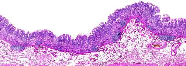
Chronic gastritis of a human, highly detailed panorama - 66 shots, 260 megapixels and after the reduced resolution. Photomicrograph as seen under the microscope, 10x zoom.

Microscope slide staining tissue biopsy for diagnosis in pathology laboratory. Staining is an auxiliary technique used in microscopy to enhance contrast in the microscopic image.

Closeup lens of a modern microscope in a research lab on a dark blue background. High resolution studio image.
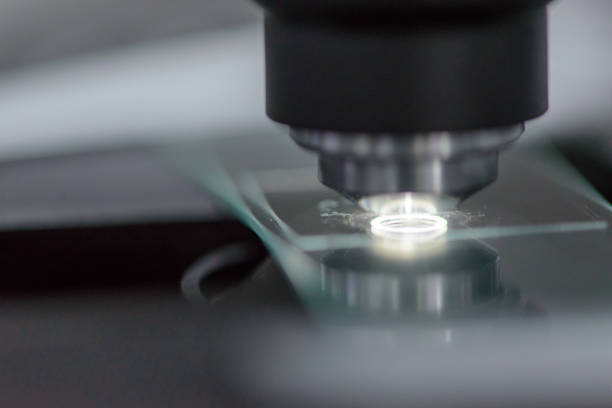
Close-up detail of a glass slide containing cancer samples with cover under the objective lens of a microscope, with bright glowing light. Science and medicine concept.
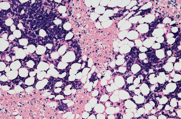
Micrograph of myeloma neoplasm bone marrow biopsy. Kappa positive insitu hybridization (ISH positive)
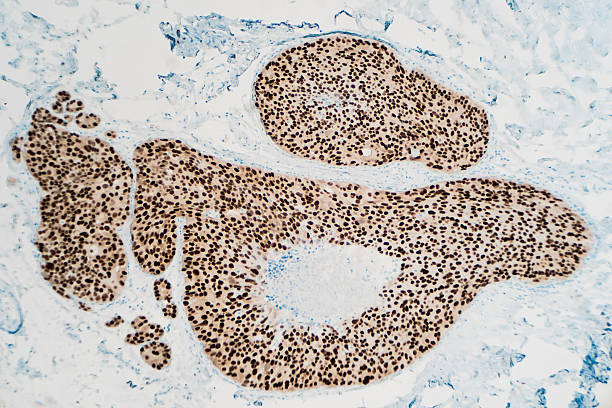
Breast cancer, ductal carcinoma in situ (DCIS): Immunohistochemistry (IHC) for estrogen receptor (ER) shows positive nuclear staining (photographed and uploaded by certified US surgical pathologist).

Hepatology concept vector for medical website, landing page. Concept of hepatitis A, B, C, D, cirrhosis, world hepatitis day. Tiny doctors treat the liver.

Culture medium bacteria colonies gram stain microscopic 100x image of Escherichia coli, also known as E. coli, is a Gram-negative, facultative anaerobic, rod-shaped, coliform bacteria.
© 2025 iStockphoto LP. The iStock design is a trademark of iStockphoto LP. Browse millions of high-quality stock photos, illustrations, and videos.
Do Not Sell or Share