Images
Pectoral Girdle Pictures, Images and Stock Photos
Browse 20,900+ pectoral girdle stock photos and images available, or search for pectoral girdle bone to find more great stock photos and pictures.
Most popular
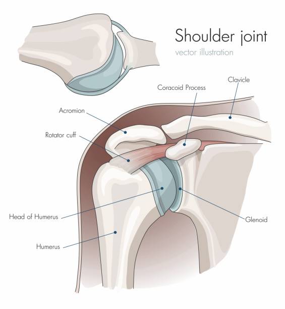
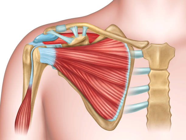
Anatomy of the shoulder joint, labeled. Vector Illustration

Closeup front angle view of a late 50's female doctor examining a male athlete with some shoulder pain. She's rotating and twisting his shoulder joint and trying to determine which tendons have been damaged. The patient has a painful grimace on his face.
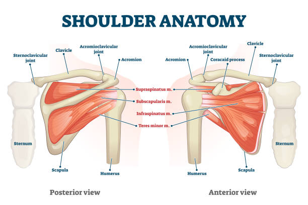
Shoulder anatomy vector illustration. Labeled inner skeleton and muscle structure scheme. Physiological educational posterior or anterior view with bones titles and location. Healthy organ description

Musculus triceps brachii 3d medical vector illustration on white background, human arm from behind eps 10

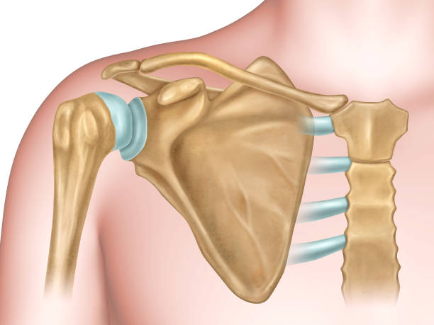
Illustration showing the bones of the shoulder

Pork Roast with Roasted Vegetables on a Platter -Photographed on Hasselblad H3D2-39mb Camera

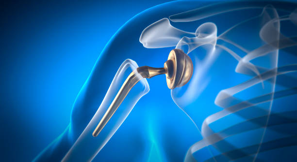
human shoulder replacement,Shoulder arthroplasty.
Muscles of shoulder and arm 3d medical vector illustration on white background eps 10


Pork Roast with Roasted Vegetables on a Platter -Photographed on Hasselblad H3D2-39mb Camera
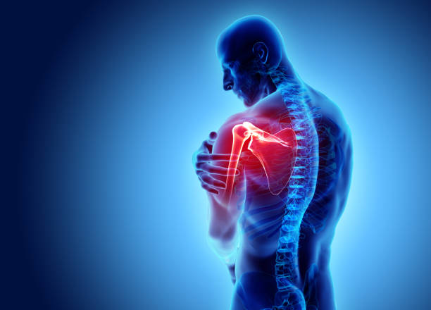
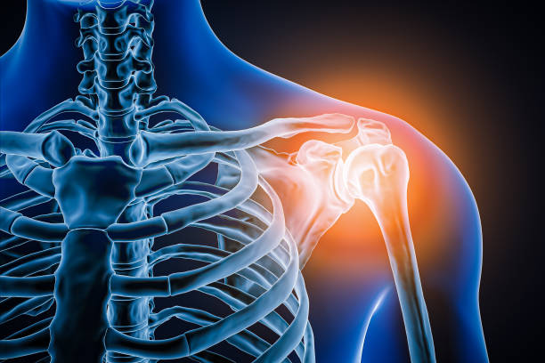
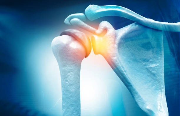
3D illustration, shoulder painful skeleton x-ray, medical concept.

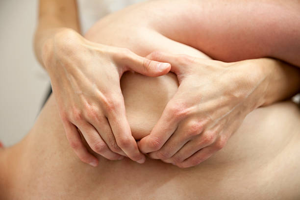
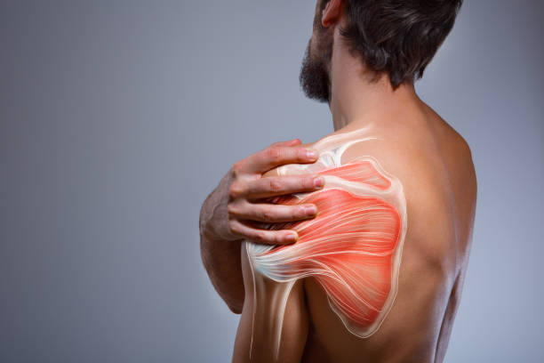
Osteopath or Physiotherapist massaging her patients scapula
High resolution 3D rendering of an injured shoulder in pain. Composite image of x-ray includes clipping plane for background change.

Sporty muscular with ponytail doing stretching workout of the shoulders, blades in sport bra, holding the neck the hand on black studio background with empty copy space. Back view. Black and white
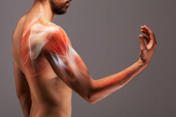
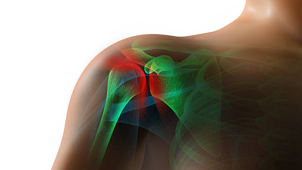
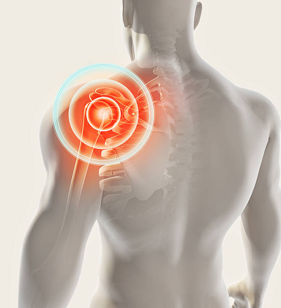
Anatomy of Shoulder , rotator cuff tear, Shoulder pain. 3d illustration


Shoulder dislocation types. Arm injury, upper arm bone pops out of the cup-shaped socket of shoulder blade. Glenohumeral joint dislocation. Flat vector illustration
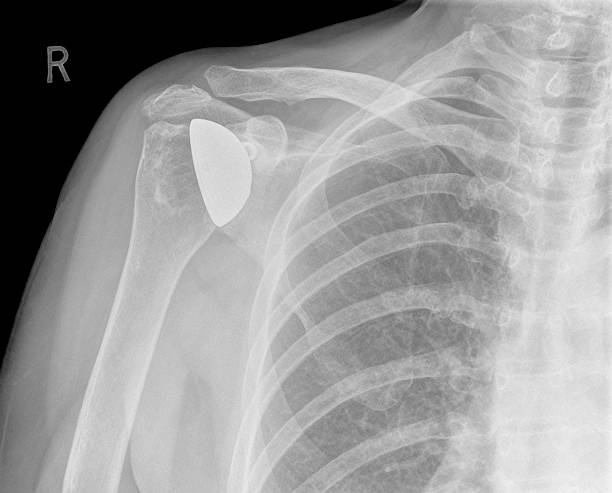
The shoulder joint has been replaced at surgery for arthritis as seen on this digital AP x-ray of the right shoulder. The articular surface of the humerus or arm bone has been replaced.
Shoulder joint replacement implant - X-ray view - Medically 3D illustration with blue background


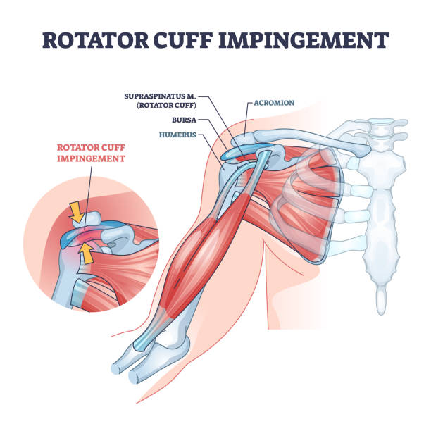
Rotator cuff impingement and anatomical shoulder muscle outline diagram. Labeled educational muscular and skeletal description with injury example vector illustration. Supraspinatus body part location
Anterior view of the shoulder anatomy. Digital illustration.

Joint implantation set. Injured or distracted human joint replaced with artificial prosthesis made of titanium. Osteoporosis and osteoarthritis treatment surgery. Flat vector illustration

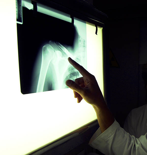
Doctors Hand pointing at an x ray of a dislocated shoulder with a stethoscope in his hand

Victorian engraving of the human Ligaments of the Shoulder and Elbow
Examinating an x-ray at hospital
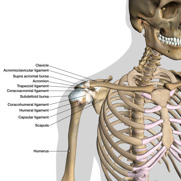
Labeled human anatomy diagram of man's shoulder ligaments, connective tissue and skeleton anterior view on a white background.
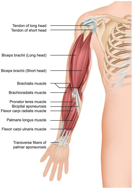
Arm muscle anatomy 3d medical vector illustration forearm eps 10
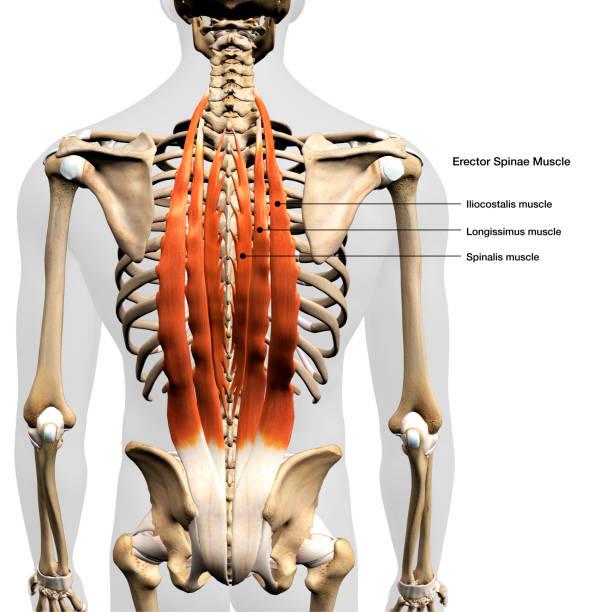
Labeled human anatomy Erector Spinae back muscles in isolation on the skeletal system from a posterior view on a white background. 3D rendering.
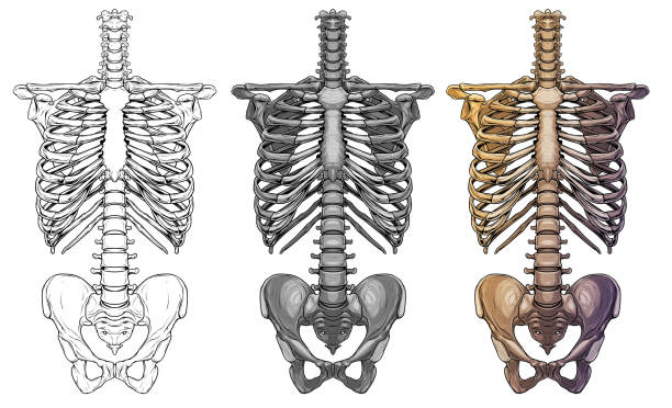
Graphic detailed black and white colorful human skeleton thorax bones. Isolated on white background. Vector icon set.
Anterior view of the shoulder anatomy. digital illustration.


Intervertebral spine hernia, pain between the shoulder blades, woman suffering from backache at home, spinal disc disease, painful area highlighted in red
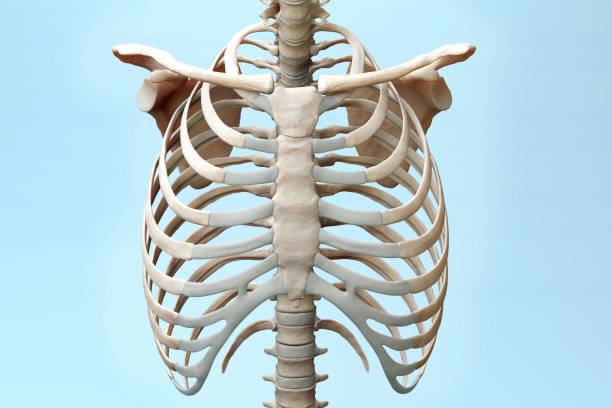
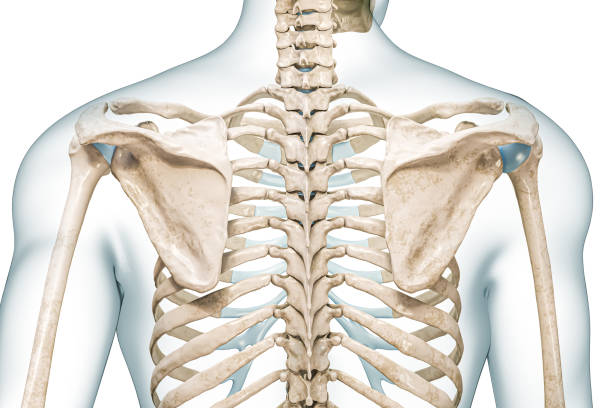
Front Torso Skeleton 3d Illustration on Blue Background

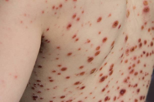
Chickenpox is an infectious disease. It causes a spotty itchy rash on the body. Some people have a few spots while others can have a lot. The spots fill with a fluid under the top layer of the skin creating bubble-like sac vesicles. These blisters eventually turn into scabs.
3D illustration, shoulder painful skeleton x-ray, medical concept.
The doctor holds in his hand a medical x-ray of a dislocated humerus and a fractured collarbone against the background of a girl patient whose shoulder hurts. Fixing bandage for the shoulder joint. Tramatology and orthopedics, close-up

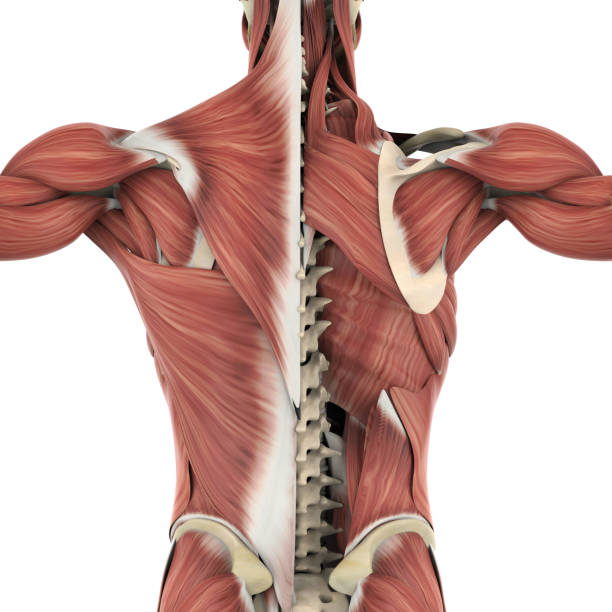
Muscles of the Back Anatomy isolated on white background. 3D render

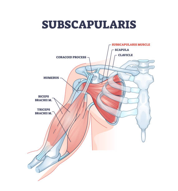
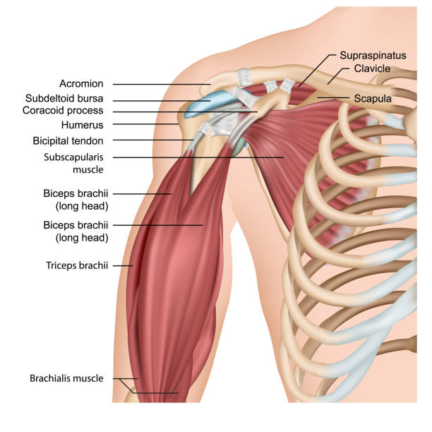
Subscapularis muscle and human shoulder inner skeletal part outline diagram. Labeled educational body scheme with medical physiology description vector illustration. Detailed anatomical bone graphic.
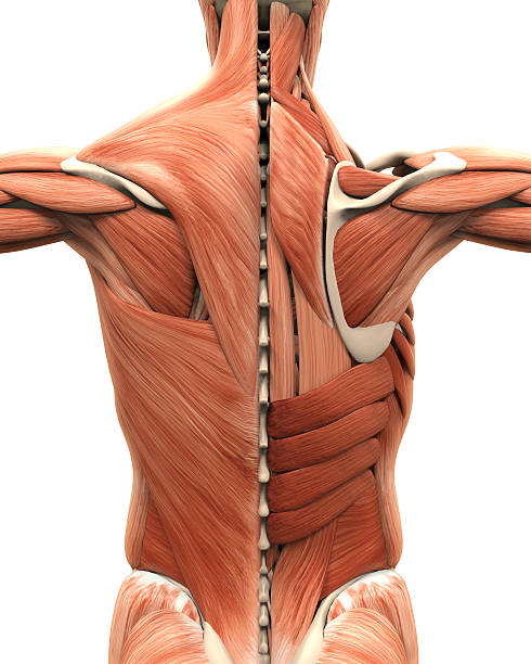
Muscular Anatomy of the Back Illustration. 3D render
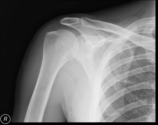
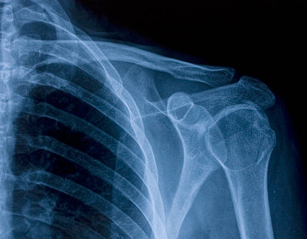
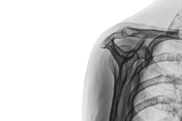
Right shoulder radiograph, AP (anteroposterior) view. No visible pathologic changes in bone structures, and no calcification in the rotator cuff. There are visible effects of the pleurectomy.

Shoulder joint replacement implant - X-ray view - Medically 3D illustration with blue background

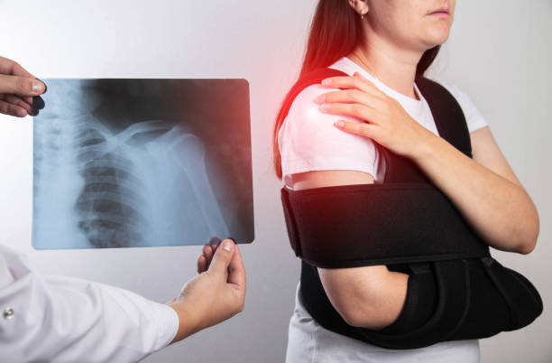
Woman wearing arm sling and looking at X-ray image. Female suffering from shoulder, clavicle, acromion fracture, strain. Health care, injury diagnostics concept. High quality photo


Sternocleidomastoid cervical muscle labeled educational anatomical scheme. Head rotation and neck flexion medical explanation vector illustration. Human model with skeleton structure and location.
Next