Photomicrograph of
Browse 10+ photomicrograph of penicillium stock photos and images available, or start a new search to explore more stock photos and images.
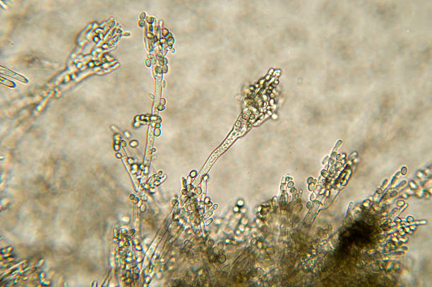
High scale magnification of a Penicillium species fungus. At the end of the long conidiophore are the phialides that form the spores. Some species of Penicillium are used in cheese making, others produce the antibiotic penicillin.

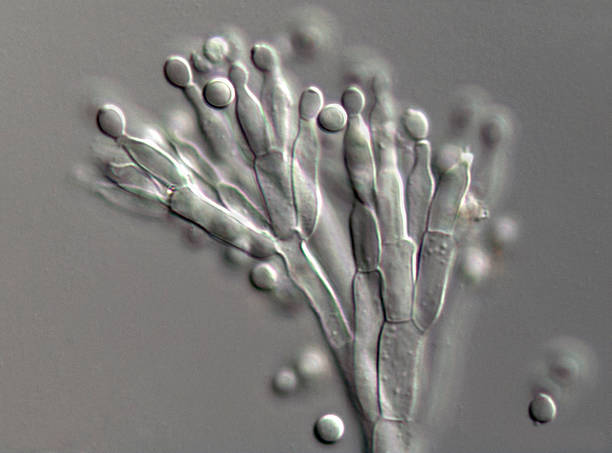
A high scale magnification photograph of the conidia and conidiophores of a Penicillium species fungus. At the end of the long conidiophore are the phialides that form the spores. Some species of Penicillium are used in cheese making, others produce the antibiotic penicillin. The photograph was taken with a light microscope fitted with Nomarski differential interference contrast optics that give high contrast at maximum aperture for optimal resolution.

"Microscopic photo of a professionally prepared slide demonstrating the cellular structure of the object.NOTE: Shallow DOF, uneven focus and chromatic aberration are inherent in microscopy, and what appears as dust is actually in the sample.See all my"

"Magnification 100X.Biological trinocular compound microscope with plan achromatic objectives and Canon 5D Mark II used for this photograph. Brightfield illumination. Note aa uneven focus, and very shallow depth of field is characteristic to photomicrography."

"Microscopic photo of a professionally prepared slide demonstrating the cellular structure of the object.NOTE: Shallow DOF, uneven focus and chromatic aberration are inherent in microscopy, and what appears as dust is actually in the sample.See all my"

"Microscopic photo of a professionally prepared slide demonstrating the cellular structure of the object.NOTE: Shallow DOF, uneven focus and chromatic aberration are inherent in microscopy, and what appears as dust is actually in the sample.See all my"

A high scale magnification photograph of the conidia and conidiophores of a Penicillium species fungus. At the end of the long conidiophore are the phialides that form the spores. Some species of Penicillium are used in cheese making, others produce the antibiotic penicillin. The photograph was taken with a light microscope fitted with Nomarski differential interference contrast optics that give high contrast at maximum aperture for optimal resolution.
© 2025 iStockphoto LP. The iStock design is a trademark of iStockphoto LP. Browse millions of high-quality stock photos, illustrations, and videos.
Do Not Sell or Share


