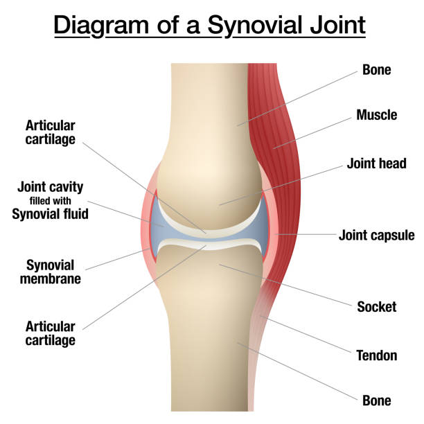
Synovial joint anatomy. Movements between the adjacent bones. Articular capsule and joint cavity filled with synovial fluid. Ligaments and cartilage in the human body flat vector medical illustration
Browse 120+ synovial membrane stock photos and images available, or search for abstract science backgrounds to find more great stock photos and pictures.
Synovial joint anatomy. Movements between the adjacent bones. Articular capsule and joint cavity filled with synovial fluid. Ligaments and cartilage in the human body flat vector medical illustration
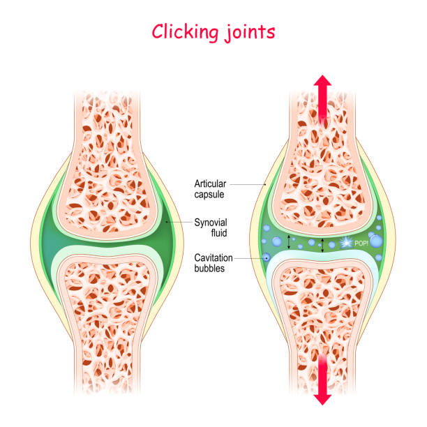
Joints and popping sound. Physiological Mechanism of cavitation. manipulate the joint we will get sound this is rapid movement of gas bubbles within the synovial solution. Cracking joint

Normal synovial joint anatomy. Healthy joint detailed illustration.

Synovial joint anatomy. joint capsule with synovial fluid and membrane. Vector illustration

Ganglion Cyst of the Wrist and Hand. Synovial cyst or a Gideon s Disease, or a Bible Cyst, or a Bible Bump. Vector illustration

Knee bursae. synovial pockets or sacs that surround the knee joint cavity. Synovial joint anatomy. Frontal and side view of human knee joint. Vector illustration

In four representations standing and isolated on white background.

Synovial joint anatomy abstract bright design. Healthy joint detailed illustration.

Vector illustration of the joint anatomy. bone. vector
Synovial bursa of the human knee. Synovial bursa is a sac filled with lubricating fluid, located between tissues such as bone, muscle, tendons, and skin, that decreases rubbing.

3D Isometric Flat Vector Conceptual Illustration of Types Of Synovial Joints, Labeled Anatomy Infographic

Hemarthrosis disease medical poster, bleeding inside of the synovial joints. A physical examination in an orthopedic clinic. Blood in joint cavity, hemophilia pain, swelling in leg vector illustration

Synovial joint anatomy. Movements between the adjacent bones. Articular capsule and joint cavity filled with synovial fluid. Ligaments and cartilage in the human body flat vector medical illustration

synovitis. Close-up. comparison and difference between a healthy joint and a joint with inflammation of the synovial membrane. Signs and symptoms of the disease

Synovial joint anatomy. Movements between the adjacent bones. Articular capsule and joint cavity filled with synovial fluid. Ligaments and cartilage in the human body flat vector medical illustration

Bursitis. inflammation of bursae (synovial fluid). Prepatellar bursitis (housemaid's knee) and Infrapatellar bursitis

Normal synovial joint anatomy. Healthy joint detailed illustration.

Rheumatoid arthritis on fingers medical vector illustration on a white background

Types of arthritis Knee and a healthy knee vector illustration on a white background

Light micrograph of a synovial membrane (innermost part of the joint capsule). It delimits the joint cavity of the diarthrosis. A band of synovial cells is concentrated in the vicinity of the lumen of the joint cavity, adopting an appearance reminiscent of a lining epithelium; however, these synovial cells are connective tissue cells that form a discontinuous lining. Deeper is a space occupied by loose connective tissue, with numerous adipocytes. On top, the articular cavity is lined by transition between synovial membrane and articular cartilage can be seen.

Synovitis. Human Knee joint anatomy. Healthy joint, and Inflammation of the synovial membrane. front view. anterior aspects. Vector illustration

synovitis of a Knee. Close-up of normal joint, and knee with inflammation of the synovial membrane. Signs and symptoms of the disease. side view of human knee joint. Vector illustration

Rheumatoid arthritis illustration. Auto immune disease, inflammatory type of arthritis

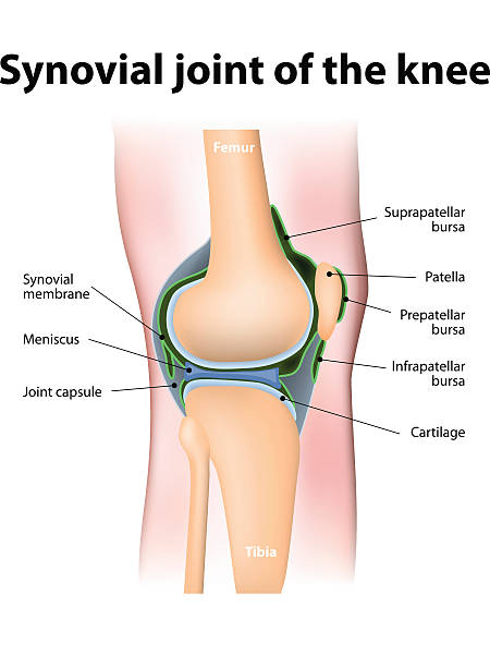
The knee joint joins the thigh with the leg and consists of two articulations: one between the femur and tibia, and one between the femur and patella. It is the largest joint in the human body. Vector graphic.

Rheumatoid arthritis and Osteoarthritis. Comparison between a Arthrosis and RA joint. Diagnostic images to aid patient and doctor. Realistic transparent blue joint on dark background. Vector illustration like X-ray image

Synovial joint anatomy. Movements between the adjacent bones. Articular capsule and joint cavity filled with synovial fluid. Ligaments and cartilage in the human body, skeleton vector illustration
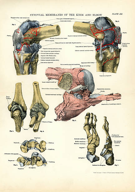
Victorian engraving of the human Synovial membranes of the Knee and Elbow

3D Isometric Flat Vector Illustration of Types Of Synovial Joints, Labeled Anatomy Infographic. Item 3

3D Isometric Flat Vector Illustration of Types Of Synovial Joints, Labeled Anatomy Infographic. Item 1

Illustration of a View of the ligaments of the inner side of the foot joints

3D Isometric Flat Vector Illustration of Types Of Synovial Joints, Labeled Anatomy Infographic. Item 2

3D Isometric Flat Vector Illustration of Types Of Synovial Joints, Labeled Anatomy Infographic. Item 4

3D Isometric Flat Vector Illustration of Types Of Synovial Joints, Labeled Anatomy Infographic. Item 6

3D Isometric Flat Vector Illustration of Types Of Synovial Joints, Labeled Anatomy Infographic. Item 5

Vector illustration of a healthy synovial joint. Anatomic poster

Synovial joint anatomy. Movements between the adjacent bones. Articular capsule and joint cavity filled with synovial fluid. Ligaments and cartilage in the human body, skeleton vector illustration

Synovial membrane with small blood vessels covering the fat pad and anterior cruciate ligament (ACL) in the intercondylar space of the left knee.

Illustration of aLigaments of the foot joints

Rheumatoid arthritis is a chronic inflammatory disorder that can affect more than just your joints. Info graphic vector.

Rheumatoid arthritis. A comparison between a healthy knee and joint with Bone erosion, Cartilage wears, Reduced joint space and Swollen inflamed synovial membrane. Diagnostic images to aid patient and doctor. Front view of human knee with glowing effect. Realistic transparent blue joint on dark background. Vector illustration like X-ray image

Illustration of a View of the ligaments of the inner side of the foot joints

Illustration of a Frontal cut of the tibio-tarsal joint

Illustration of a Interosseous ligaments and synovial membranes of the articulations of the bones of the foot

Illustration of aLigaments of the foot joints

Light microscope micrograph of a human synovial membrane (innermost part of the joint capsule). It delimits the joint cavity of the diarthrosis. A band of synovial cells is concentrated in the vicinity of the lumen of the joint cavity, adopting an appearance reminiscent of a lining epithelium; however, these synovial cells are connective tissue cells that form a discontinuous lining. Below is a loose connective tissue with abundant blood vessels and adipose tissue. On the right is the fibrous connective tissue of the fibrous capsule.
Rheumatoid arthritis line black icon. Vector isolated button. Editable stroke.
Rheumatoid arthritis color line icon. Diseases of the spine. Vertebrology. Vector isolated element. Editable stroke.
Rheumatoid arthritis color line icon. Autoimmune diseases. Vector isolated element. Editable stroke.

Joint anatomy symbol set. Colorful illustrations

Human synovial membrane showing large chronic inflammatory infiltrates (chronic synovitis) and synovial cell hyperplasia, a morphology that can be seen in rheumatoid arthritis

From First Book on Analytic, Physiology and Hygiene 1872