Images
X Ray Pelvis Pictures, Images and Stock Photos
Browse 5,000+ x ray pelvis stock photos and images available, or start a new search to explore more stock photos and images.
Most popular
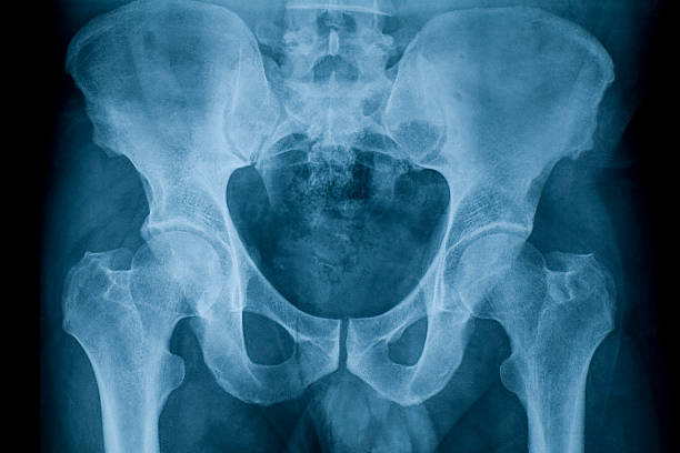

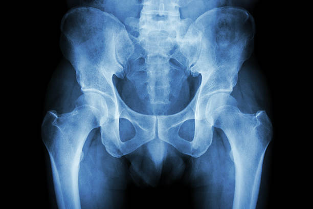
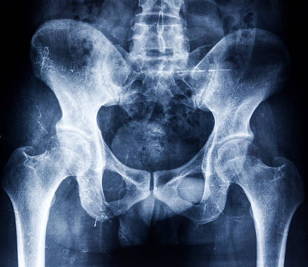
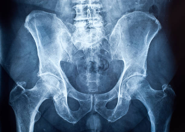
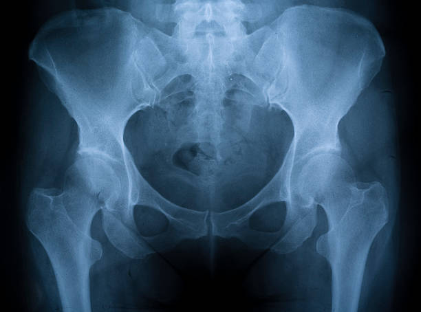
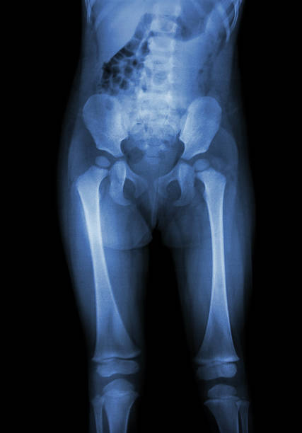
X-ray close up pelvic bone and hip joint

Medical team examining X-ray image of a pelvis in the hospital.
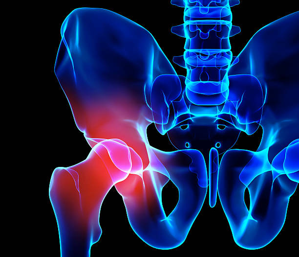
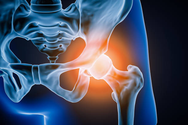
3D illustration, hip painful skeleton x-ray, medical concept.
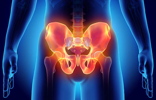
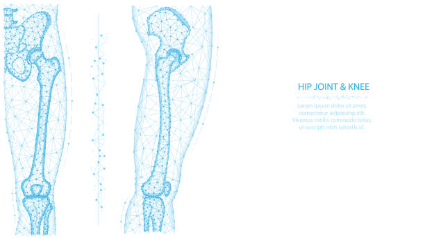
3D illustration of Hip Skeleton, medical concept.

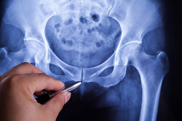
Pretty young woman doctor watching x-ray image of human pelvis....pointing at it
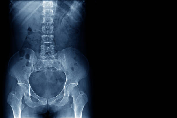
Xray of an adult female pelvis taken at a hospital. The female is suffering from pelvic bone misalignment and a rotation of the L3 vertebrae. Minor arthritis is beginning to show in the left hip socket/joint. No damage (cracks/fractures) to any bones or vertebrae found; diagnosis tentatively points to lower back sprain.
Kidney stone ( renal stone , renal calculi ) ( film x-ray KUB ( Kidney - Ureter - Bladder ) show left renal stone )

Mid adult doctor examining X-ray image of a pelvis in the hospital.


Doctors sitting around the table and interpreting x-ray image
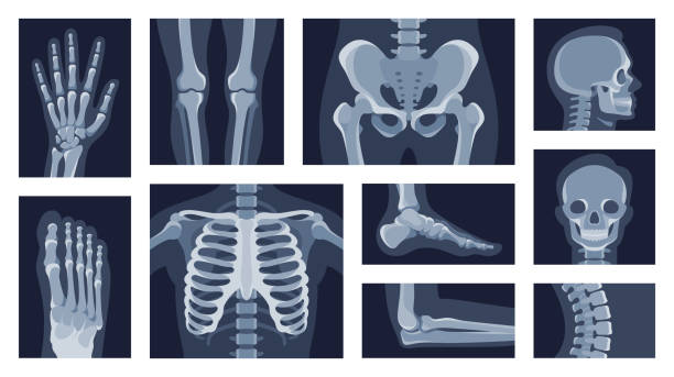
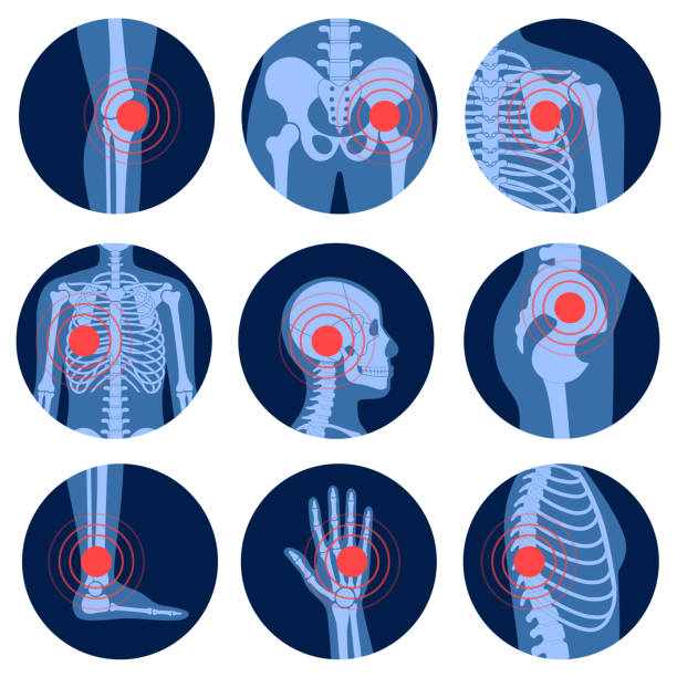
Collection different human body parts roentgen pictures vector flat illustration. Set x rays shot of head, hands, legs, torso, chest, hip, elbow, knee, spine skeleton character isolated on white
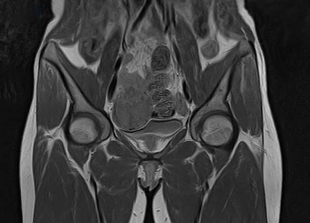
magnetic resonance imaging or mri scan report of spinal cord or lumbar bulging, x-ray, Lumbar Spine (Low Back), lower disc bone problem, bulging disc, herniated disc, slipped disc scan
X-ray image of a female pelvis with a lumbar vertebra fracture. Human skeleton. Front view.

Female doctor showing to female senior patient an x-ray on the tablet

lumbar vertebrae x-ray image by mri on digital tablet screen with medical team for consult.

Walking man painful hip joint with blue background- x-ray illustration
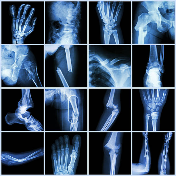
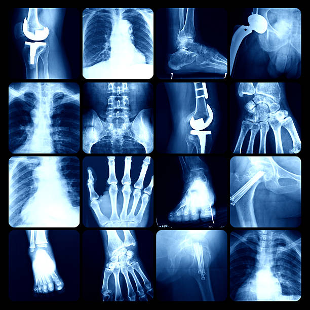
Collection X-ray multiple bone fracture (finger,spine,wrist,hip,leg,clavicle,ankle,elbow,arm,foot)

Shot of a young doctor using a digital tablet during a consultation with a senior woman

pelvis x-ray image of after childbirth women shows distasis of simphysis pubis called symphysiolysis

Human man skeleton pain, fracture or inflammation, parts of male body on x ray view. Vector isolated flat illustration of skull and bones on reontgen. Medical, educational or science banner.
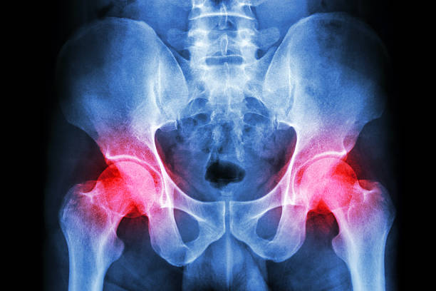
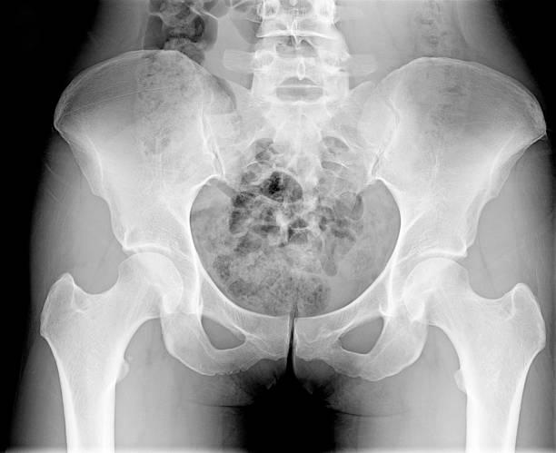
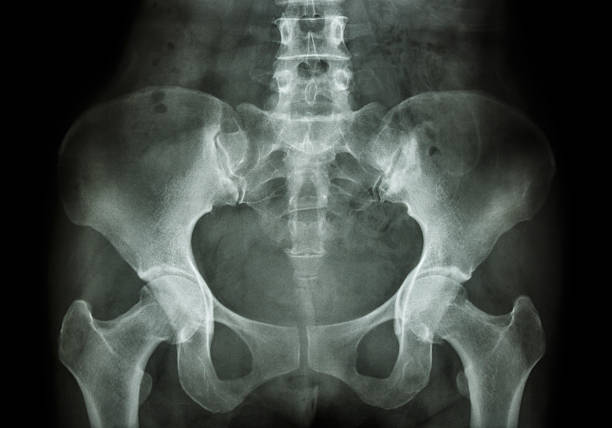
film x-ray human's pelvis and arthritis at both hip joint (Gout , Rheumatoid)

Set line icons of orthopedics isolated on white. Vector illustration


Mid adult doctor examining X-ray image of a pelvis in the hospital.


X-ray shots of human body. Cartoon vector illustration. X-rays of pelvis, chest, knees, feet in black background. Skeleton, X-ray, bone roentgen, medicine, health, research concept for banner design
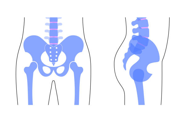
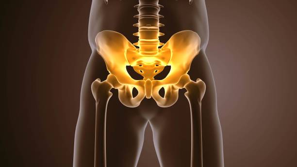
Human pelvis anatomy. Main pelvis bones - sacrum, ilium, coccyx, femur. Front and side view. Vector illustration isolated on white background. skeleton silhouette. Medical, educational banner
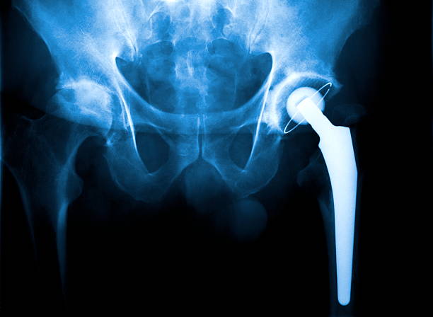
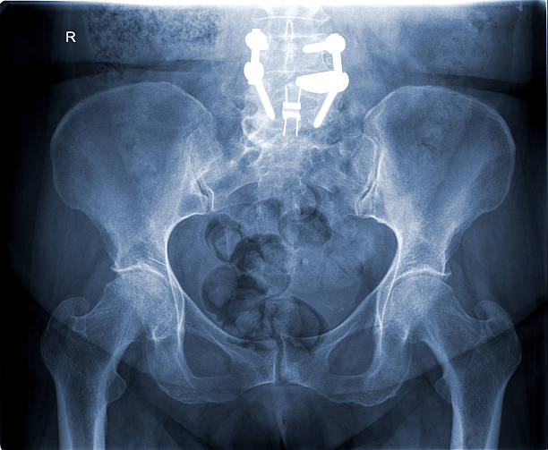
Real X-ray of a Arthroplasty of the hip joint. (Abbreviation: HTP or H-TEP)
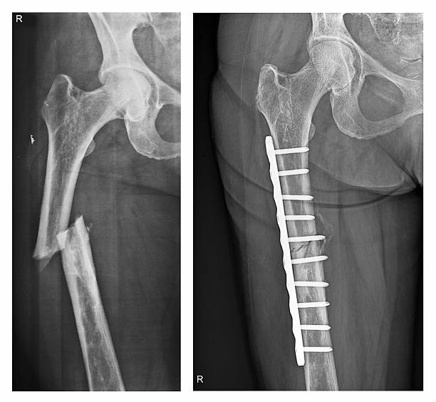
X-ray of a femur fracture (broken thigh bone) before and after surgery. The fracture of the femoral shaft is not quite healed after the surgery.

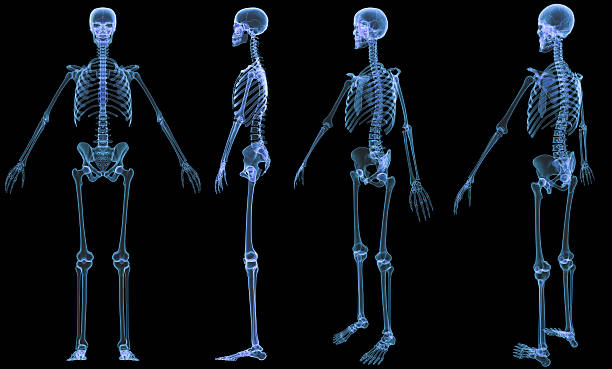
Digital medical illustration: X-ray of human skeleton.
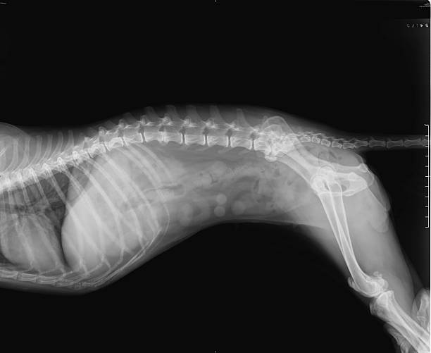
Negative X-Ray of spinal column, chest, abdomen, pelvis and thighbone of a female 16 years old small dog

Shot of a doctor using a digital tablet during a consultation with his patient


Delve into the complexities of an arthritic sacroiliac joint with this detailed anatomy concept image. Explore the skeletal structure and learn about the implications of inflammation and disease in this critical area of the body. Ideal for medical education, presentations, and healthcare-related content.
Human joints and body parts bones sketch icons. Vector isolated set of spine pelvis, shoulder scapula or elbow, leg knee and foot ankle, arm and hand wrist with fingers for medical anatomy or surgery


Hip joint pain, woman suffering from osteoarthritis at home, health problems concept

concept of health care technology, parts of skeleton in anatomical science

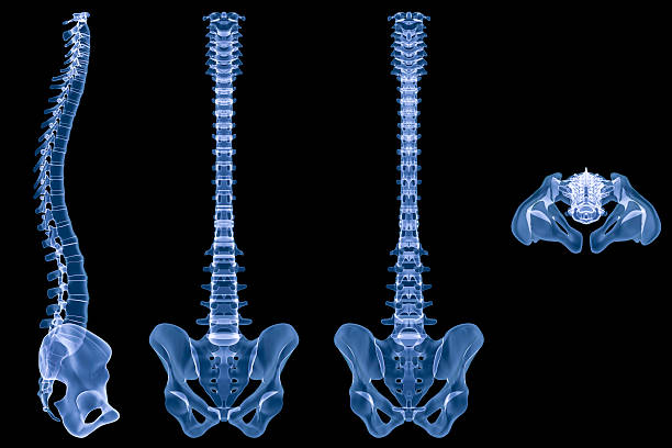
Digital medical illustration: X-ray human spine. 4 views included:
Magnetic Resonance Imaging (MRI) of uterine with contrast
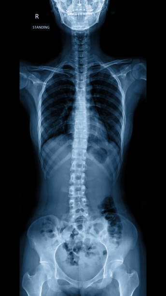
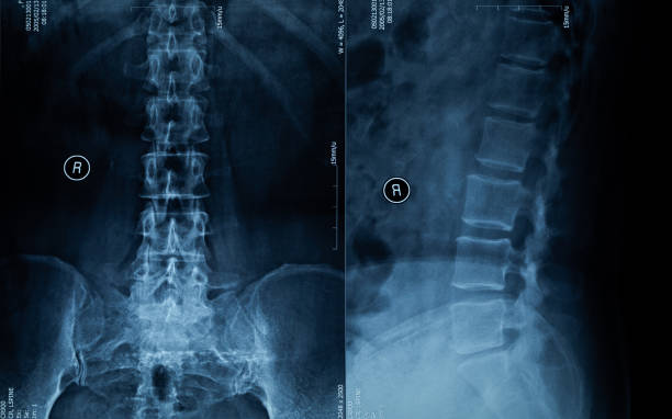
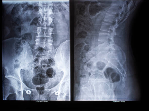
x-ray of whole spine showing straight spine of all parts cervical, thoracic and lumbar spine.

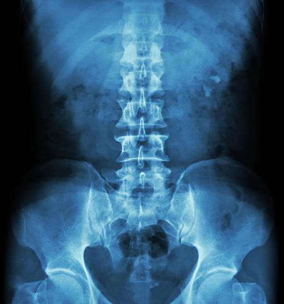
Human spine in x-ray, on gray background. The lumbar spine is highlighted by red colour.
Next